In this series of posts we are going to examine these seemly simple questions:
- What is the goal and the purpose of testing of unknowns generally? How do we best design a test for marijuana?
- How is most marijuana testing conducted in the United States?
- What is microscopic morphological examination? Is it a “good” test?
- What is the modified Duquenois-Levine test? Is it a “good” test?
- What is Thin Layer Chromatography? Is it a “good” test?
- Is the combination of all three tests create a “good” testing scheme?
- Is there a better way to test for marijuana?
Part 3: What is microscopic morphological examination? Is it a “good” test?
Microscopic morphological examination
What is it?
The microscopic morphological examination in short is an exercise of botanical identification using a microscope.
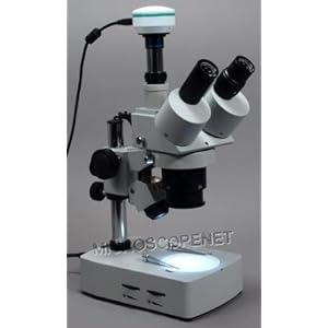
Mechanically how is it preformed?
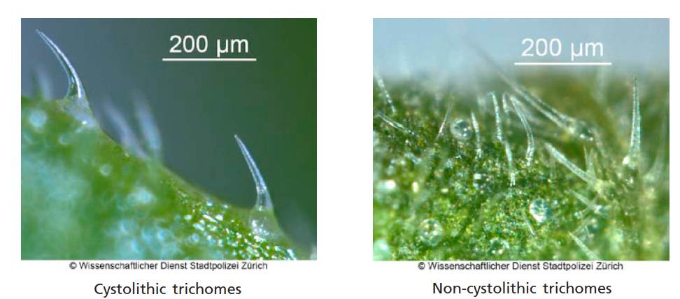
A very small amount of the dried unknown is selected. This becomes the sample. The sample is placed on a microscopic slide. A drop or two of water is then added to the slide. The slides are examined at varying levels of magnification and under different light conditions. What the analyst is looking for is two distinct morphological features. They are looking for microscopic “hairs” on the unknown. These are cystolithic hairs and glandular hairs. Cystolithic hairs are often likened to like little bear claws in their appearance.
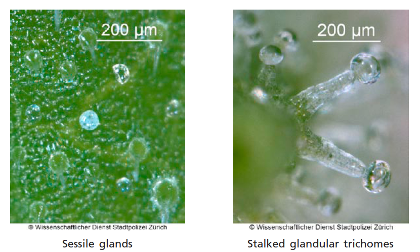
The second type of hair is called a glandular hair. These are frequently remarked as looking like mushrooms.
Some techniques call for the use of hydrochloric acid after they look for these hairs. A few drops of HCL are added by the analyst. The analyst then looks to see if there is some unspecified effervescence under the light of the microscope.
How is the typical crime laboratory analyst trained to conduct this form of testing?
The question becomes what experience level in botany and taxonomy and microscopy does the analyst truly have? Very few undergraduate programs exist in botany in the United States. Most analysts have on the job training where another person who likewise have no formal training in botany or taxonomy that instructs them. It also involves the use of a microscope. Formal training in microscopy is required in order to use a microscope properly and to properly interpret what the human eye sees through various powers and lighting conditions of the microscope. At the end of the in-house training, the typical analyst cannot typically express the family, the genus and the species that is “marijuana” or at what power and under what lighting conditions they saw the morphological characteristics.
This sample that is examined under the microscope is then discarded. All future or additional testing is conducted on totally different samples from the unknown.
Is this a verifiable test?
It potentially is. There is a device that can be linked to the microscope to take pictures of what the analyst thinks he or she sees. This is called a photomicrograph. In fact the pictures above come from just such a microscope that is equipped with one. A digital camera attached to a microscope is very commonly used in science. They are very moderately priced. As they are digital cameras, the cost of production and printing and data storage is negligible. It is frequently used in other types of comparative examinations such as some higher levels of forensic firearm or toolmark identification. I know of no laboratory in the United States that relies upon microscopic morphological examination that uses modern technology and produces photomicrographs. In fact, few, if any, crime laboratories use the ACE-V (Analysis, Comparison, Evaluation, and Verification) technique that one would find in fingerprint identification using a stereo-microscope and a double check in real time by a fellow bench analyst. In essence, the unknown is checked one time, by one person with no double check by another, and nothing is produced that proves that the analysis was done or that the features that are reported as present were in fact objectively present. There is no proof.
Is there empirical validity studies that prove that this is a specific and confirmatory test?
No. There are no empirical and robust validation studies that conclude that this form of microscopic morphological examination even when the two botanical features (cystolithic and glandular hairs) are objectively present yield a valid opinion that the plant examined is definitely contains THC. There are no studies that say the two features means that there is THC present. In fact, what studies that are out there conclude that this form of morphological examination using a microscope is perhaps not even selective. In the original studies by Nakamura, he indicated that cystoliths of various types are found in the leaves of a number of dicots. (497). He also indicated that the presence of cystoliths is not diagnostic for a family, let alone a genus of plants. (497) Nakamura specifically noted that cystoliths are found on a great number of plants including but not limited to: hops plants (500), oregano (500), lemon thyme (501), silver thyme (501), and rosemary (501). Nakamua he specifically noted 63 “representative” species in 13 plant genera that contain cystoliths in table 5 of his article (501) Nakumura indicated that he made NO attempt to prepare a comprehensive listing of species bearing cystolith hairs similar to those found in cannabis “because of the sheer enormity of the task to examine 31,874+ dicotyledons.” (500). For instance, in one genus found in Table 5 of his article, the Loasa, he specifically noted 9 species that had cystoliths; however, he went on to say that there were actually some 80 species of that genus known to have similar hairs. (501). He fully acknowledged that his listing was not comprehensive. So it is accurate and very fair to say that the 63 “representative” species that have cystoliths that were noted by Nakamura in Table 5 of his article are not an exhaustive list. Other studies agree that at least 6 other substances also have hairs that contain these two features (cystolithic and glandular hairs). In terms of the additional step of adding HCL to the sample and examining for effervescence under the light of the microscope, it is quite clear that other substances can produce the same effervescence when a few drops of hydrochloric acid are added to them. For example nettles and catnip do exactly that.
Some folks maintain and testify under oath every day in the United States that this unverified microscopic morphological examination is diagnostic of identification of THC presence in an unknown. There is no scientific support for this type of testimony.
One thing that every analyst should agree with is that simply because these hairs are present and if they conduct HCL addition and if there is effervescence that does not mean that the unknown contains THC. This is why they have to do additional testing, meaning the modified Duquenois-Levine and Thin Layer Chromatography testing.
What is frequently not part of any morphological examination for cannabis is what botanists have noted to be other features consistent with cannabis. The simplified examination for the typical forensic science identification is purposefully designed to make this examination and conclusions from it easier to perform by non-botanists. As with everything in life, the more criterion attached to qualify something the least likely there will be a qualification. The examination of cannabis and especially a morphological examination by untrained botanists should not be made easy. All of the features that are known to be diagnostic by the world of botany should be used not simply the easy ones. For example, botanists have noted that that cannabis has sessile glands as well as containing serrated edges of the leaves and compound palmate structure meaning several leaflets arise from the same point. The addition of all of the known morphological features known to true botanists as diagnostic of cannabis would make this examination more robust and the result more selective than the simplistic examination that now permeates the forensic science world.


Melissa says:
“I know of no laboratory in the United States that relies upon microscopic morphological examination that uses modern technology and produces photomicrographs.”
Georgia.
Justin J. McShane says:
Thank you. If you can give details, I would be happy to amend, acknowledge and recognize what I think is good and best forensic practices.