The following is a brief summary of the modern explanation of gunshot wounds according to the proponents of interpreting gunshot wounds (GSW). After you read this, you draw your own conclusions as to whether or not this is empirical or more towards the subjective or non-validated.
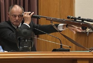
Gunshot Wounds: A summary form a pathology textbook
In general, when a person is shot, the injury sustained will result in a temporary wound cavity that is produced due to cavitation, which occurs when a body moves so quickly in a liquid that the liquid detaches from the body surface. This cavity will only exist for a short period of time after the penetration of the projectile. The size and seriousness of the wound cavity will depend on the amount of energy transmitted by the gun, which is dependent on the length of the barrel; the longer the barrel, the greater velocity.
There are four categories of wounds: (1) contact wounds, which can be hard, loose, angled or incomplete; (2) near contact; (3) intermediate; and (4) distant.
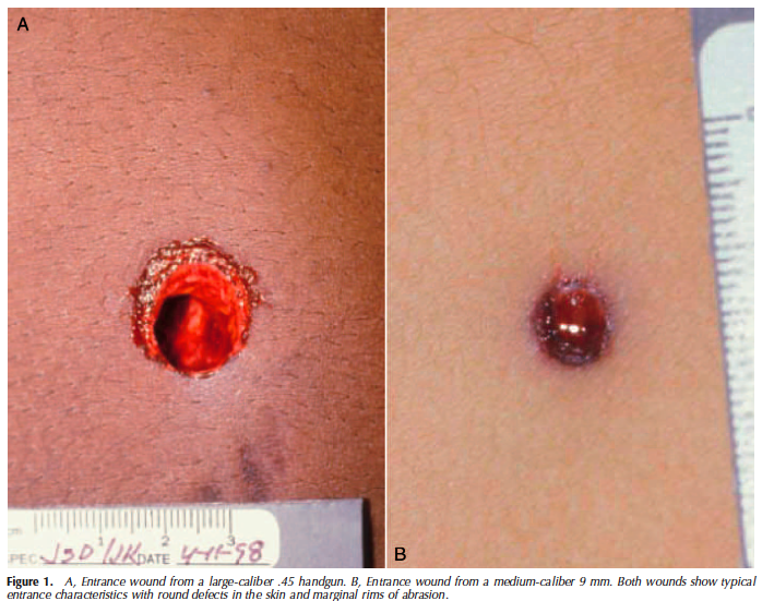
When the wound is a contact wound the muzzle of the gun is placed up against the body at the time of discharge. When this occurs gas, soot, metallic particles, vaporized metal, primer residue, and powder particles can be found in the wound track. Hard contact wounds result from the muzzle being held very tightly against the skin and will create an indent. Due to the closeness of the muzzle to the skin, all the materials from the muzzle will be left directly in the wound which leaves very little external evidence on the skin. If a proper autopsy was conducted soot and un-burnt particles would be found in the wound track. During the autopsy soot could easily be distinguishable from dried blood as soot, unlike dried blood, cannot be removed by either water or hydrogen peroxide. Furthermore, if a dissecting microscope was used, not only would soot always be present in the wound, but also the powder particles left in the wound could be identified.
Where the projectile pierces skin that is tightly flexed over bone, like the skull, the wound will have a different appearance because the gas discharge, which expands between the skin and outer table of the bone, lifts the skin and causes it to “balloon out”. When the stretching of the skin exceeds the elasticity of the skin, the skin will tear. The subsequent size of the tear will depend on the caliber of the weapon used, the amount of gas produced, the firmness of the weapon held to the body, and the elasticity of the skin. This type of tear, however, can also occur when the victim is shot at an intermediate or distant range if the bullet is able to perforate the skin over a bony prominence or curved area of bone that is covered by a thin layer of tightly stretched skin.
Where gas is the cause of the tearing of the skin, however, the detail of the imprint left on the skin will depend on the amount of gas produced from the firing of the gun. The more gas produced, the harder the skin will impact against the muzzle, which results in a greater, more detailed imprint. Imprints cannot only be found on the skin but will be found in the chest and the abdominal region as well because the gas produced will expand in the visceral cavities and adjacent soft tissue causing the chest or abdominal wall to bulge out creating larger imprints which can be twice the actual size of the muzzle.
The presence of a loose contact wound suggests that the muzzle is held in very light contact with the skin as the skin is not indented by the muzzle. In this type of situation, the gas preceding the bullet and the actual bullet itself will be the cause of the indent on the skin. Any soot left on the skin can be easily swiped away but an autopsy can and will reveal particles of powder, vaporized metals, and soot deposited in the wound track, along with carbon monoxide.
In near contact wounds, the second category, clumps of unburned power can pile up on the edges of the entrance wound and on the seared zone of the skin.
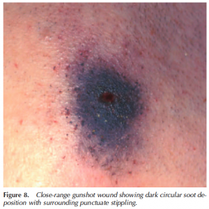
The third category, intermediate wounds, are formed when the muzzle is held away from the body at the time of discharge but is still sufficiently close enough that the powder grains from the muzzle can strike the skin and produce powder tattooing, which gets it name from the blackening of the skin around the entrance site caused from the soot. The size and density of these tattoos will depend on the caliber, barrel length, type of propellant powder, and the distance from the muzzle to the target. As the range away from the target increases, the intensity of powder blackening will decrease and the size of the soot pattern area will increase. The powder tattoo which results is unlike the soot in the loose contact wound in that it cannot be swiped away from the skin.
Finally, distant wounds, will be created when the muzzle is sufficiently far from the body so that there is neither deposition of soot nor powder tattooing in the wound track. When a person is shot from a distance, the clothing of that individual will absorb the soot and the powder, thus making it essential for the victim’s clothing to be examined during the autopsy. As the clothing absorbs most, if not all of the soot, the ability of the powder to leave a mark on the skin of the victim will depend on the nature of the material, the number of layers of cloth, and the physical form of the powder.
_______________________________
Blogger’s comments:
The problem with these types of descriptive groupings lies in the validation. These definitions and descriptions of observable phenomena sounds scientific, but is it? It sounds like something that is empirically based, but is it? How is it possibly measured? How are these conclusions validated? How can we have confidence that these categories and the underlying observations that describe them are unique as to that category and not present in the other categories? We can’t.
Some of the problem comes down to one of the very basics: documentation. Usually in autopsies, photographs are taken. Sometimes they are not taken according to basic principles of photogrammetry. The photos are taken from oblique angles instead of at 90 degrees and most of them have no scaling mechanism. This lack of meaningful photography makes it difficult to verify measurements taken, produce any sort of scale or otherwise reconstruct the wound characteristics. In addition how the investigator approaches documenting the wound can drastically change the interpretation. For example, if one were to clean the wound site or shave it with the idea of revealing it, then gun shot particles, sooting or other characteristics may be removed. In addition, some examiners still use metal rods to try to determine the “path of the bullet” by probing the wound and inserting rods through it. This will deform and alter the organic state of the wound. In the worst case scenario, it can actually create a false path through soft tissue and therefore lead to an erroneous conclusion. Rather than just relying on the typical “I know it when I see it” characteristics outlined above that is used in modern gunshot wound examination, we should use computerized tomography, fluoroscopy and even radiographs including CAT scans.
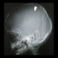
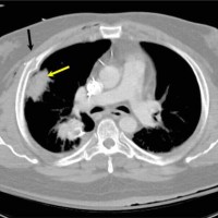
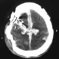
Now, after seeing these computerized tomography images, who can argue with what is more persuasive, “I know it when I see it” or the available science?



William the Coroner says:
In 10 years of practice, in a large city and smaller offices, I have never seen a CT scan of a cadaver outside of a research protocol. Plain-films should always be obtained on GSW and knife wounds, but the time and expense of getting the scanner does not justify the results.
Secondly, though soot can be removed from the skin surface by washing, stippling (by definition) canNOT be removed by washing. As it is made by gas expansion forcing the burning/unburnt powder into the skin.
To do proper range of fire testing, the laboratory must use 1. the suspect firearm 2. ammunition from the same lot number and manufacture as the shooting. Powder age, composition, barrel length, intermediate target all effect the amount of gunshot residue seen.
Justin J. McShane says:
I thank you for coming to the site and I welcome your opportunity to comment so that your point-of-view can be expressed.
I respectfully disagree.
You are willing to put a price tag on the criminal justice process and a person’s liberty. I do not enjoy the “it costs too much” argument as a justifiable excuse. Certainly, the “it takes too long” excuse is perhaps worse. With respect, do you volunteer to have yourself or a family member or friend have anything less than certainty?
Crime Lab says:
Of course there is a price tag on the criminal justice system. Does a wealthy person who can afford top of the line attorneys and private investigators not stand a better chance than poor schlub who gas a P.D? Also, our system of justice is based on guilt beyond a REASONABLE doubt, not guilt beyond anything less than certainty.
jeff howerton says:
Kudos to Crime Lab…does justice have a price, haha. Does a bum who gets shot in the alley get the same forensic workup as say if the president were assasinated???? Basically in a world with finite resources, the amount of time(and money as well) spent on the “pursuit of justice” is directly linked to the amount of people who want/demand to know the cause. So, like it or not, the amount of people who are affected by the outcome of the autopsy and findings will directly affect the means to which the coroner will have at his disposal.Sadly, no one really cares if an improper wound track angle would send the bum standing in the middle of the alley to jail vs the one at the end from an incorrect assesment.
Yuaas says:
Sir, I am confused Between the Charring and Blackning. What is the major difference between charring and blackning around the gUn Shot wounds.
IF it is then where i can find a detailed article on these two, and if not they are similer then in what sence they are one. Is there any one who can help me with authentic reasons